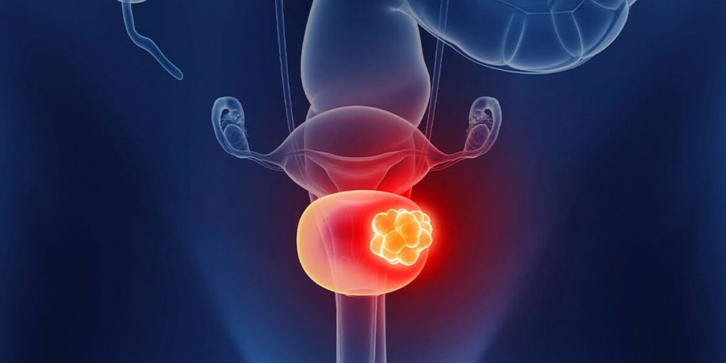
Prostate cancer is a significant health concern for many men, particularly as they age. Early detection and attentive care can make all the difference in treatment outcomes. This article delves into various aspects of prostate cancer, focusing on how to recognize early signs, the importance of regular screenings, and effective management strategies.
Prostate Cancer: What to Know
Prostate cancer develops in the prostate, a small gland that produces seminal fluid. It’s one of the most common forms of cancer among men. While the exact cause remains unclear, certain risk factors can increase vulnerability, including age, family history, and ethnicity. A significant statistic reveals that one in eight men will be diagnosed with prostate cancer in their lifetime. Early detection is crucial as it can potentially lead to more effective treatment options and better survival rates.
Recognizing Symptoms: Early Signs to Watch For
Many early stages of prostate cancer are asymptomatic, making regular medical check-ups essential. However, there are subtle signs that may indicate a problem. Common symptoms can include difficulty urinating, blood in urine or semen, persistent pain in the lower back or hips, and unexplained weight loss. Understanding these signs and discussing them with healthcare providers can facilitate early diagnosis. In many cases, men may dismiss or normalize these symptoms, which can delay the diagnosis. Awareness and vigilance are crucial in boosting the chances of timely intervention.
The Role of Screening: Importance of Regular Check-Ups
Screening for prostate cancer typically involves a blood test for prostate-specific antigen (PSA) and sometimes a digital rectal exam (DRE). The American Urological Association recommends that men discuss the risks and benefits of these tests with their healthcare providers starting at age 50 or earlier for those at higher risk. Early screening can lead to the detection of cancer before it spreads, significantly improving treatment success. Various studies have shown that men who undergo regular screenings have a higher likelihood of catching the disease in its most treatable stages.
Diagnosis and Staging: Understanding Your Options
Once prostate cancer is suspected, further diagnostic tests are essential. This may include imaging tests such as MRI or CT scans and a biopsy to confirm the presence of cancer cells. The cancer is then staged to determine how advanced it is, which is critical for planning treatment. Understanding the stage of the disease informs the possible options available, including active surveillance, surgery, radiation, or hormone therapy. Each pathway has its considerations, and engaging in thorough discussions with healthcare professionals helps men and their families make informed decisions.
Treatment Plans: Tailoring Care for Individual Needs
Prostate cancer treatment varies widely based on factors like the cancer type, stage, and the patient’s overall health. Active surveillance may be recommended if the cancer is slow-growing and not showing symptoms. On the other hand, more aggressive forms might necessitate intervention through surgery or radiation therapy. Hormonal therapy seeks to reduce levels of male hormones that encourage cancer cell growth. Each treatment has pros and cons, and discussing these thoroughly with oncologists and other specialists can help narrow down the best approach for each individual’s unique situation.
Lifestyle Changes: Supporting Health During and Post-Treatment
Embracing a healthy lifestyle can be beneficial for men both during treatment and in recovery. Diet plays a significant role; many studies suggest that diets rich in fruits, vegetables, and whole grains may contribute to better outcomes. Regular exercise can also help in managing weight and improving overall well-being. Educating oneself about prostate cancer contributes to proactive participation in one’s care. Joining support groups can provide emotional support and access to shared experiences from others in similar situations, empowering men to navigate their journeys more effectively.
The Importance of Ongoing Monitoring: Staying Proactive After Treatment
Survivorship doesn’t end with treatment; ongoing monitoring is crucial to catching any potential recurrence early. Regular follow-ups and check-ups can aid in identifying any changes or symptoms that might arise post-treatment. These appointments typically include blood tests for PSA levels and periodic imaging to ensure that any new developments are addressed promptly. Staying informed about the disease, engaging actively in follow-up care, and maintaining open lines of communication with healthcare providers significantly increase the chances of long-term survival and quality of life.
Taking proactive steps in understanding prostate cancer and actively participating in healthcare decisions can greatly influence outcomes. Awareness of the signs, the significance of regular screenings, and tailored treatment plans form the foundations of early detection and care. By keeping informed and engaged, individuals can ensure they are well-positioned to tackle prostate cancer effectively. Education about prostate health is empowering, and the journey toward awareness does not end at diagnosis; it continues long after treatment. Men and their loved ones have access to numerous resources and communities, allowing them to share experiences and support one another through this journey of survivorship.
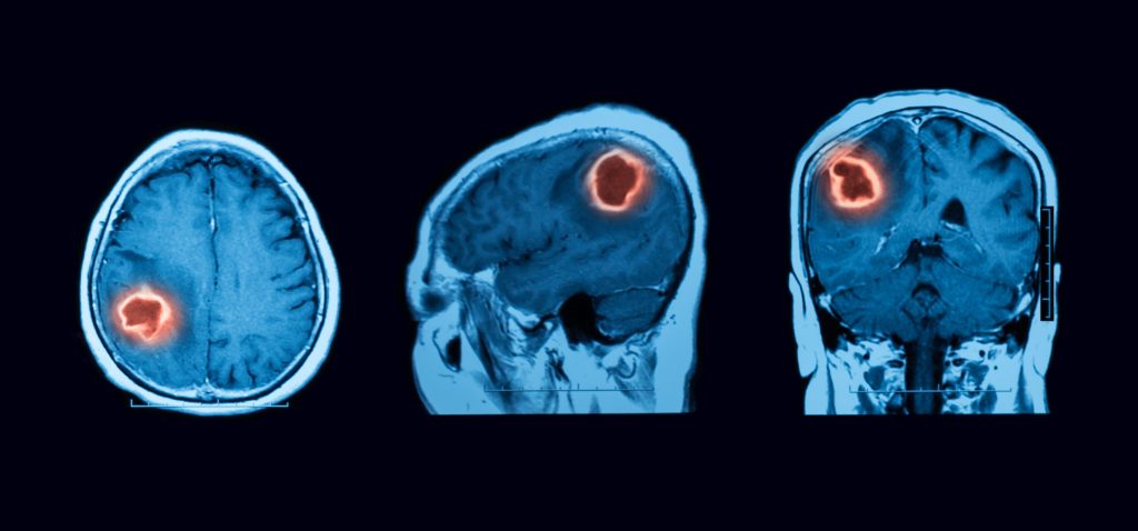
The human brain is an incredible, complex organ that controls numerous body functions, thoughts, and emotions. However, when something goes wrong, such as the development of a brain tumor, early intervention can be crucial for better outcomes. Recognizing early signs of a brain tumor and the associated symptoms is important for timely diagnosis and treatment.
One of the key indicators that something might be amiss is persistent headaches. While headaches are generally common and can stem from various benign causes like tension or dehydration, a notable shift in the nature of headaches can raise suspicion. For instance, if the headaches occur more frequently, intensify over time, or are associated with nausea and vomiting, they could indicate increased intracranial pressure due to a tumor. According to the American Brain Tumor Association, headaches affect about 50-70% of patients with brain tumors. Therefore, it’s essential to be attentive if headaches change in character or severity, particularly if they begin to disrupt daily activities or sleep.
Another prevalent symptom is changes in vision. This can present through blurred vision, double vision, or sudden loss of vision in one or both eyes. These changes occur because brain tumors can affect areas responsible for processing visual information or putting pressure on the optic nerve. The National Eye Institute reports that vision changes can occur in one out of three individuals diagnosed with a brain tumor. Consequently, situations involving unexplained changes in visual perception should not be taken lightly and may warrant further investigation.
Additionally, cognitive and behavioral changes can serve as red flags for the presence of a brain tumor. Individuals may notice memory lapses, confusion, difficulty concentrating, or personality changes. Such symptoms can manifest gradually, and family members might be the first to observe these changes. Research shows that about 40% of brain tumor patients experience cognitive changes, which could result from the tumor’s location or pressure it exerts on surrounding brain tissue. If cognitive decline becomes apparent, it is crucial to document specific changes and consult a healthcare provider for further assessment.
Seizures are another significant symptom associated with brain tumors. While many people may not connect seizures to a potential tumor, the American Academy of Neurology notes that tumors are a common cause of new-onset seizures in adults. Seizures can manifest in various ways—from convulsions to more subtle symptoms like unusual sensations or changes in behavior. Approximately 20-50% of individuals with brain tumors experience seizures at some point, thus making it essential to seek medical evaluation if seizures occur, especially if they are not part of a known diagnosis.
Motor skill and coordination changes are additional symptoms to consider. These may present as weakness, clumsiness, or difficulty with balance and coordination. When a tumor affects areas of the brain responsible for movement and coordination, individuals may find that simple tasks become challenging. For instance, difficulty holding or gripping objects can be a sign that something is amiss, potentially linked to a brain tumor. According to a study published in the Journal of Neurosurgery, motor deficits are often seen in 30-40% of brain tumor patients, highlighting the importance of closely monitoring any changes in physical abilities.
Lastly, changes in sensory perception can occur in individuals with brain tumors. This can include altered sensation such as numbness, tingling, or the inability to feel touch in specific body areas. These symptoms may arise due to a tumor pressing on or damaging nerves responsible for sensation. As with other symptoms, this should not be overlooked, especially when it presents alongside other warning signs. Acknowledging these changes can be essential for timely diagnosis and treatment.
Recognizing early signs of a brain tumor can make a significant difference in treatment outcomes. Although many of the symptoms mentioned above can stem from various non-cancerous conditions, being proactive and vigilant about changes in health is vital. If someone experiences multiple symptoms persistently or to an unusual degree, seeking medical advice is crucial. Healthcare professionals can provide assessments and initiate necessary imaging tests, such as MRI or CT scans, to check for abnormalities.
Maintaining open lines of communication about potential symptoms with healthcare providers, along with regular health check-ups, helps in keeping track of one’s health. Awareness of these symptoms and the desire to seek assistance can set a pathway for better outcomes. Timing is crucial as brain tumors can differ significantly in type and growth rates, meaning that early detection can provide a broader array of treatment options.
In conclusion, recognizing early signs of a brain tumor and understanding their associated symptoms is essential for timely intervention and better health outcomes. Pay attention to major changes in headaches, vision, cognitive functions, motor skills, and sensory perception, as they might signal a need for further investigation. This proactive approach can empower individuals to take control of their health and enable healthcare providers to act decisively, potentially leading to more favorable outcomes. Listening to one’s body, observing changes, and consulting with medical professionals is essential in navigating any health concerns with diligence and care.
Blood in the urine isn’t something to brush off, yet many people delay a checkup hoping it will fade on its own. Bladder cancer can start quietly, hiding behind symptoms that resemble common infections or routine aging. Knowing the early warning signs and risk factors can turn uncertainty into a clear plan for action. This guide walks you through what to watch for and when to call a clinician, in straightforward, calm terms.
Outline: – Why bladder symptoms matter and a quick tour of how the bladder works – Hematuria and urinary changes as early signals – Non-urinary symptoms and red flags not to ignore – Risk factors, look-alikes, and what else could explain symptoms – When to seek medical care, what tests may be used, and how to prepare
Why Bladder Symptoms Matter: A Quick Tour of the Organ and What Signals Mean
The bladder is a muscular reservoir that collects urine from the kidneys and releases it through the urethra. Its inner lining, called the urothelium, is constantly exposed to chemicals filtered from the bloodstream. Because of this close contact, troublesome cells can sometimes arise in the lining. Early-stage disease is often confined to the inner layer, while more advanced disease can grow into the bladder wall and beyond. Recognizing symptoms promptly can influence the path forward, helping clinicians find issues when they are more manageable.
Symptoms matter because they are the body’s early alert system. While many bladder complaints turn out to be benign, overlooking repeating or unusual patterns can delay answers. Painless blood in the urine, for example, may appear suddenly, disappear, and return weeks later. Such a stop-start pattern can feel reassuring when it fades, yet it still warrants evaluation. Even subtle urinary changes, like going more often or a new nighttime pattern, can be meaningful when they persist. The aim is not to cause worry but to encourage thoughtful attention and timely conversations with a healthcare professional.
Here’s why taking note of bladder changes is worthwhile: – The bladder’s lining can bleed from irritation, infections, stones, or tumors; sorting these out requires testing. – Some concerning conditions cause no pain at first, so absence of discomfort is not a reliable green light. – Intermittent symptoms still count; a single episode of visible blood is a reason to schedule an appointment. – Early evaluation often leads to simpler treatments and clearer choices.
A helpful habit is to jot down what you notice: color changes in urine, frequency, urgency, burning, or pelvic pressure. Include dates and any related triggers, such as strenuous exercise, new medications, or dehydration. This small record can guide the discussion with a clinician and make the most of the visit. Most importantly, do not self-diagnose; overlapping conditions are common, and appropriate testing is the safest path to clarity. Hematuria and Urinary Changes: The Most Common Early Signals
Blood in the urine, known as hematuria, is the hallmark symptom linked to bladder tumors. It can be visible (turning urine pink, red, or cola-colored) or microscopic (detected only on a urine test). Visible blood may appear without pain and may stop and start over time. Clots can occasionally form and look like small strings or jelly-like pieces. Because exercise, infections, stones, and certain medications can also lead to blood in the urine, it is essential to let a professional sort out the cause rather than assuming it is minor.
Other urinary changes can accompany or precede hematuria. These include a newfound urge to urinate, going more frequently, difficulty starting the stream, burning or stinging, and a sensation of not emptying fully. Nighttime urination that is new for you may also be notable. None of these are proof of cancer, but a cluster of persistent urinary changes—especially when paired with any episode of blood—raises the need for timely evaluation. Comparisons help: a short, sharp burning sensation with fever and foul-smelling urine often points toward infection; a waxing and waning colicky pain that radiates to the groin can suggest a stone. Bladder tumors, by contrast, may cause painless bleeding or a steady escalation of irritative symptoms without the classic peaks of stone pain.
When urine looks different, note: – Color: pink, orange-tinged, red, brown, or cola-like – Presence of clots: small strings or gelatinous pieces – Timing: at the start, throughout, or at the end of the stream – Frequency patterns: sudden increases by day or night
Questions to ask yourself before an appointment: – Did I recently start a new medication, vitamin, or strenuous exercise plan? – Do symptoms improve with hydration or worsen after certain activities? – Is there fever, flank pain, or foul odor suggesting infection? – How many episodes of visible blood have occurred and over what time span?
Even a single episode of visible blood is a cue to schedule a visit. Persistent microscopic blood found during a routine check is also worth investigating. Early conversations do not commit you to invasive procedures; they open the door to a stepwise evaluation tailored to what you are experiencing.
Beyond the Bathroom: Non-Urinary Symptoms and Red Flags
While bladder-related symptoms center on urine changes, the body sometimes sends less obvious signals. Pelvic discomfort or a sense of pressure can develop when the bladder lining is irritated or when growths occupy space. Low back or flank pain may occur if urine flow is affected, particularly when swelling or blockage develops near where the kidneys drain. Unintended weight loss, loss of appetite, or persistent fatigue can reflect the strain of ongoing illness and deserve attention when they persist without a clear explanation. These signals are nonspecific, yet their value lies in the pattern: when combined with urinary changes or blood in the urine, they strengthen the case for evaluation.
Red flags that merit prompt medical contact include: – Visible blood in the urine with clots or difficulty passing urine – Increasing pelvic pain or a new constant ache above the pubic bone – Back or side pain with fever, chills, or nausea – New swelling in the legs or feet, which can signal impaired drainage – Repeated “urinary infections” that clear briefly and then recur – Unexplained fatigue, weight loss, or anemia noted on routine labs
It helps to compare possible causes. Infections often bring burning, urgency, fever, and rapid improvement after a short course of appropriate treatment. Kidney stones commonly cause severe, wave-like pain and sometimes blood, but the pain typically surges and falls as the stone moves. Bladder tumors can be quieter, featuring painless bleeding or irritation that lingers rather than spikes. No single symptom tells the whole story; the combination, duration, and response to initial care provide the clues. Because some serious conditions begin subtly, erring on the side of an early conversation with a clinician is a practical, low-risk decision.
If any symptom disrupts your routine or triggers worry—particularly visible blood—set up an appointment rather than waiting for a next flare. Most evaluations start with simple tests, and many people leave with reassuring answers. If something requires attention, finding it sooner keeps more options on the table.
Risk Factors, Look-Alikes, and What Else Could Explain Symptoms
Understanding risk factors can sharpen your radar without creating alarm. Tobacco exposure, current or past, is a major contributor to bladder lining irritation. Age plays a role; risk increases as decades pass, and many diagnoses occur after midlife. Occupational exposure to certain dyes, solvents, or combustion byproducts has been linked to higher risk, particularly with long-term contact. Chronic bladder irritation—from longstanding catheters, repeated infections, or stones—can also nudge the lining toward abnormal changes. Prior pelvic radiation or some chemotherapy agents have been associated with later bladder issues. Family history is less common as a direct driver but can combine with other factors to shift risk.
Key factors to consider: – Tobacco exposure over years, including past use – Long-term contact with specific industrial chemicals or fumes – Repeated bladder irritation from infections, stones, or catheters – Prior pelvic radiation or exposure to certain anticancer drugs – Advancing age and male sex, though all adults can be affected
Many bladder symptoms have look-alikes. Urinary infections can mirror urgency, burning, and frequency, often with fever or foul-smelling urine. Kidney or ureteral stones can cause blood in the urine and sharp flank pain. In many men, prostate enlargement leads to weak stream, nighttime trips, and incomplete emptying. Gynecologic sources, such as vaginal bleeding mistaken for urinary bleeding, can complicate the picture. Interstitial cystitis and overactive bladder create urgency and frequency without infection. Dehydration, strenuous exercise, and certain foods or supplements can temporarily shift urine color as well.
Common mimics to keep in mind: – Urinary infection: burning, urgency, fever, quick response to treatment – Stones: wave-like flank pain, occasional nausea, microscopic or visible blood – Prostate-related symptoms: hesitancy, weak stream, nocturia – Gynecologic bleeding: spotting or cycles overlapping with urination – Bladder pain syndromes: urgency and pressure without infection
Sorting these possibilities relies on testing rather than guesswork. A straightforward plan—urinalysis, culture if infection is suspected, and imaging when indicated—often separates short-term, self-limited issues from conditions that require targeted care. Risk reduction is always worthwhile: staying hydrated, avoiding tobacco, using protective gear when working with chemicals, and seeking care early for recurrent infections can all support bladder health.
When to Seek Medical Care, What to Expect, and Practical Next Steps
If you see blood in your urine—even once—schedule a medical appointment. Seek urgent or emergency care if you cannot pass urine, pass large clots, or have severe pain with fever. For symptoms like persistent urgency, frequency, mild pelvic pressure, or microscopic blood found on a routine test, plan to see a clinician within a reasonable timeframe rather than waiting months. Early evaluation does not automatically mean invasive procedures; instead, expect a stepwise approach that matches your symptoms and risk profile.
Common first steps may include: – Urinalysis to check for blood, protein, and signs of infection – Urine culture if infection is suspected – Blood tests to look at kidney function and anemia – Imaging, such as ultrasound or a specialized CT scan, to view the kidneys, ureters, and bladder – Cystoscopy, a direct look at the bladder lining with a thin scope, when warranted by symptoms or test results
Preparing for your visit can make it more productive: – Keep a symptom diary with dates, urine color changes, frequency, pain scores, and potential triggers – List medications, vitamins, and supplements, including start dates – Note any tobacco exposure, past or present, and workplace chemical contact – Bring prior test results or imaging reports if you have them – Write down questions about next steps, timelines, and follow-up
Understanding possible outcomes reduces anxiety. Many people discover a benign explanation—such as a temporary infection or a small stone—that resolves with focused care. If the evaluation identifies a bladder growth, your clinician will discuss options based on location, size, and depth, often starting with removal or sampling through the urethra. Follow-up schedules are common after any concerning finding, because the bladder lining can develop changes over time. The practical takeaway is simple: noticing and acting on early signs places you in a stronger position, regardless of the cause. Conclusion: Stay observant, document what you experience, and reach out promptly when something is new, persistent, or worrisome. Thoughtful attention today supports informed choices tomorrow.
Colon inflammation can upend routines, but clear guidance turns confusion into practical steps. This guide explains genetic risk for Crohn’s, the lived sensations of an ulcerative colitis flare, and foods that often soothe during tough days. Expect plain language, careful data, and practical tips grounded in clinical guidance without hype. Read on to feel more prepared for your next conversation with a healthcare professional.
Outline and Why This Guide Matters
Colon inflammation shows up in different ways, but two conditions are especially common: Crohn’s disease and ulcerative colitis. Both are forms of inflammatory bowel disease, and both can affect daily life in practical, sometimes surprising ways—what you eat, how you plan your day, even how you sleep. This guide gives you a grounded roadmap so that terms like “flare,” “low-residue,” or “genetic risk” stop feeling mysterious and start feeling manageable. Consider it a field manual: factual, calm, and immediately useful.
Here’s the outline we’ll follow, so you can scan ahead or read straight through depending on your question of the day: – Section 1 (you’re here): Why this topic matters, how the guide is organized, and what you can expect to learn. – Section 2: Chances of developing Crohn’s disease if a parent has it, explained with absolute versus relative risk, genetic signals, and real-world modifiers like smoking. – Section 3: What an ulcerative colitis flare often feels like—from bathroom urgency and cramps to fatigue and the mental load—plus warning signs that call for timely care. – Section 4: What foods are commonly better tolerated during a flare, how to build plates with gentle ingredients, and a sample 3-day menu to reduce guesswork. – Section 5: A practical wrap‑up with quick steps you can use tomorrow, and ways to partner with your care team without feeling overwhelmed.
Why this matters now: research keeps refining our understanding of risk and symptom patterns, but what people need most is translation—clear numbers, relatable descriptions, and food strategies that respect individual differences. That’s our approach. We’ll keep the tone steady and avoid sweeping promises. Think of the pages ahead as a map you can fold into a pocket: not flashy, but reliable when the trail gets rocky.
Chances of Getting Crohn’s Disease If a Parent Has It
When a parent has Crohn’s disease, risk does rise for their children, but context helps. In many populations, the lifetime chance of Crohn’s in the general public is low—often well under 1%. Having a first‑degree relative with Crohn’s increases that absolute risk into the low single digits for many families, commonly cited in the neighborhood of 2–9% over a lifetime. If both parents live with inflammatory bowel disease, estimates are notably higher, with reports around 30% for offspring in some cohorts. These figures vary by ancestry, environment, and the specific mix of genes a family carries.
It’s useful to separate relative risk from absolute risk. Compared with a very small baseline, relatives can have a many‑fold increase, yet the actual number across a lifetime may still be modest. Researchers have identified genetic variants—such as changes in immune‑related pathways—that raise susceptibility, but no single gene “guarantees” Crohn’s. Twin studies show higher concordance among identical twins than fraternal twins, which supports a genetic contribution, but the mismatch within identical pairs also proves that environment and the microbiome matter.
Several modifiable and non‑modifiable factors can tilt risk up or down: – Family history: strongest known predictor, especially in first‑degree relatives. – Ancestry: some groups, including certain European and Middle Eastern ancestries, report higher prevalence. – Smoking: associated with increased risk of Crohn’s and more complicated disease courses. – Early‑life exposures: antibiotics in infancy, urban living, and reduced early microbial diversity are under study as potential contributors. – Gut microbiome: patterns of microbial imbalance may precede inflammation for some individuals.
For families planning ahead, a few practical points can be grounding. First, routine screening colonoscopies for asymptomatic children of parents with Crohn’s are not typically recommended solely on the basis of family history; discussions focus on symptoms if and when they appear. Second, general health choices—avoiding smoking, maintaining regular physical activity, and adopting a balanced diet—support overall gut and immune health even if they do not eliminate risk. Finally, staying alert to early, persistent digestive symptoms (unintended weight loss, ongoing abdominal pain, prolonged diarrhea, or blood in stool) enables prompt evaluation and, when needed, earlier intervention. The takeaway: a parent’s diagnosis raises the odds, but most children of parents with Crohn’s still do not develop the condition, and day‑to‑day choices plus timely medical attention can meaningfully influence the journey.
What Does an Ulcerative Colitis Flare Up Feel Like
Ulcerative colitis primarily inflames the lining of the colon and rectum, so flares tend to broadcast themselves through the bathroom. Many people describe an urgent need to go with little warning, often several times a day, sometimes waking them at night. Stools may be loose, with visible blood or mucus. Cramping pain—commonly a steady ache that sharpens before a bowel movement—often eases a bit afterward, only to return as the colon continues to spasm. Tenesmus, the sensation of needing to pass stool even when the rectum is nearly empty, can feel like a stubborn doorbell that won’t stop ringing.
Beyond the gut, a flare can feel like a dimmer switch turning down your energy. Fatigue is common, fed by poor sleep, fluid losses, mild anemia, or the constant vigilance that comes with unpredictable urgency. Low‑grade fevers may appear, along with decreased appetite and a slow creep of weight loss if the flare lingers. Some experience extra‑intestinal symptoms—achy joints, tender skin nodules, or eye irritation—that seem disconnected from the colon but are linked by the immune system’s reach. Emotionally, flares can feel isolating: plans get cross‑checked against restroom maps, and even a short commute feels longer when every traffic light sparks a negotiation with your gut.
Not all flares announce themselves at the same volume. Mild episodes may add a couple of urgent trips with a smudge of blood. Moderate flares can mean frequent stools, more blood, and escalating cramps that color your day. Severe flares may bring near‑constant urgency, marked bleeding, persistent pain, and dehydration. Warning signs that call for prompt medical input include: – Passing blood with nearly every stool, especially if clots are present. – Fever that doesn’t settle, signs of dehydration, or dizziness. – New, severe abdominal pain that does not improve. – Inability to keep up with fluids or food for more than a day.
When describing your symptoms to a clinician, concrete details help: how many trips per day, how much blood (streaks, spoonfuls, or more), nighttime awakenings, and pain patterns before and after bowel movements. Think of it as building a precise weather report for your colon; the more accurate the forecast, the better the treatment plan and the quicker the path toward calmer days.
What Foods to Eat During a Colitis Flare Up
During an ulcerative colitis flare, the goal is to lower mechanical and chemical irritation while still nourishing your body. Many people do better, at least temporarily, with a low‑residue approach: fewer insoluble fibers that scrape and stimulate, gentler textures, and modest portions eaten more often. The keyword is “tolerance”—there is no single plate that works for everyone, but certain patterns are repeatedly reported as easier on an inflamed colon.
Consider leaning on these building blocks: – Hydration and electrolytes: water, broths, and oral rehydration solutions; add a pinch of salt and a little sugar to water during heavier losses. – Carbohydrates with lower insoluble fiber: white rice, plain pasta, sourdough or white toast, rice noodles, saltines. – Soluble‑fiber foods that form soothing gels: oatmeal cooked soft, ripe bananas, applesauce, canned peaches. – Gentle proteins: poached chicken or turkey, flaky white fish, eggs, silken tofu; prepare by steaming, baking, or poaching rather than frying. – Low‑lactose or lactose‑free dairy choices if dairy worsens symptoms: lactose‑free milk, aged cheeses in small amounts, or fortified plant milks. – Fats in small amounts: olive oil or avocado oil for cooking; avoid heavy, greasy meals.
Common triggers to approach cautiously during a flare include raw tough greens, corn, whole nuts and seeds, popcorn, spicy chilies, alcohol, high‑caffeine drinks, sugar alcohols, and fried foods. Some people identify onions, garlic, or carbonated beverages as irritants. If iron supplements upset your stomach, ask about gentler formulations or timing adjustments, since anemia can complicate recovery.
Here’s a sample three‑day, flare‑friendly sketch you can adapt: – Day 1: Breakfast—oatmeal cooked soft with mashed ripe banana; Lunch—white rice with poached chicken and peeled steamed carrots; Snack—applesauce; Dinner—baked cod with mashed potatoes (no skins) and zucchini cooked until tender. – Day 2: Breakfast—white toast with scrambled eggs; Lunch—rice noodles in mild chicken broth with tofu; Snack—ripe banana; Dinner—turkey meatballs in simple tomato‑free gravy over rice, canned peaches for dessert. – Day 3: Breakfast—cream of rice cereal with lactose‑free milk; Lunch—plain pasta with olive oil and finely cooked spinach (if tolerated); Snack—saltines and a soft cheese slice; Dinner—steamed fish, polenta, and peeled, cooked squash.
Portion size matters: smaller, more frequent meals can reduce stretch and spasm in an inflamed colon. Keep a brief food and symptom log for a week or two; patterns often appear quickly and help you personalize the plan. As symptoms ease, reintroduce a wider variety of fruits, vegetables, and whole grains gradually, starting with well‑cooked, peeled, or blended options. If weight loss or food fear creeps in, a registered dietitian experienced in inflammatory bowel disease can help rebuild variety and confidence without reigniting symptoms.
Putting It All Together: A Practical Plan and Supportive Conclusion
Living with a personal or family connection to inflammatory bowel disease is a long project, but it need not be a lonely one. The numbers around Crohn’s risk can feel abstract until you translate them into action: absolute risk is typically low even when a parent has the condition, and everyday choices add up. For ulcerative colitis, understanding what a flare can feel like lets you prepare without bracing in fear. Food strategies aren’t about perfection; they’re about stacking gentle options while your colon asks for quiet.
Here is a compact plan you can tailor: – Map your baseline: record average stools per day, pain levels, and any blood when you feel well, so changes are easier to spot. – Build a flare kit: oral rehydration packets or a homemade mix, soft snacks, spare undergarments, and moist wipes, kept discreetly in a bag or desk. – Set thresholds: for example, call your clinician if you exceed a set number of stools per day, if bleeding increases, or if fevers persist beyond 24–48 hours. – Support the fundamentals: sleep, movement, and stress‑reduction practices—short walks, paced breathing, or brief mindfulness sessions—can dampen the body’s alarm system. – Mind known modifiers: if you smoke, seek support to stop; if certain medications upset your gut, ask about alternatives or protective strategies.
As symptoms settle, widen your diet methodically and celebrate small wins, like tolerating a new fruit or adding a cooked green. For families where a parent has Crohn’s, keep the conversation open with children at a developmentally appropriate level: honest, reassuring, and focused on what to do—not what to fear. If a new digestive pattern persists, early evaluation can shorten the path to relief and reduce complications. And remember, community matters: local support groups and reputable online communities can turn hard‑won tips into shared knowledge.
Conclusion: You now have a clear view of how family history influences Crohn’s risk, what an ulcerative colitis flare may feel like, and how to steady your diet during turbulent days. Use this as a living document—annotate, adjust, and revisit. With steady information, a few practical tools, and an ongoing partnership with your care team, you can navigate colon inflammation with more clarity, less guesswork, and a growing sense of control.
Jaw bone loss can derail implant plans, loosen teeth, and chip away at everyday confidence. This guide unpacks why bone thins, what modern dentistry can do, and how smart nutrition supports recovery. We break down costs with plain-language ranges to help you budget with clarity. If you want practical steps and science-backed tips, you’re in the right place.
Why Bone Loss Matters: Introduction and Outline
Bone might seem like a silent partner in your smile, but it is the foundation that keeps teeth and dental implants stable. When the jawbone thins—whether after a tooth is removed, because of gum disease, or due to systemic factors—teeth can loosen, bite forces shift, and implant plans may need a rethink. The good news is that modern dentistry offers predictable solutions, and daily habits, including nutrition, can support the biology that rebuilds and preserves bone. This introduction sets the stage and maps the path you will follow in the sections ahead.
Here is the outline you will see unpacked in detail:
– Not Enough Bone for Dental Implant: causes, diagnostics, and surgical strategies to create or find stable bone. – Dental Implants with Bone Loss Cost: what drives fees, typical ranges, and smart budgeting tips without cutting corners. – How to Fix Teeth with Bone Loss: non-surgical and surgical gum therapies, stabilization options, and timelines for healing. – Vitamins That Support Jaw Bone Health: how nutrients interact with bone cells, realistic targets, and food-first strategies. – Putting It All Together: prevention, maintenance, and when to move from plan to action.
Why this matters now: after a tooth is removed, the ridge naturally shrinks. Research shows substantial changes in the first year, often 25–50% reduction in ridge width if a socket is left empty, with the most rapid loss occurring in the first few months. Periodontal disease compounds the problem by eroding both the gum attachment and the bone around roots. And while implants can restore function and appearance, they require sufficient bone volume and density to succeed. Understanding the biology allows you to choose treatments that work with, not against, your body.
Throughout this article you will find clear explanations, realistic timeframes, and examples that compare options. When numbers are given (for costs or timelines), treat them as planning tools rather than promises; individual cases vary by health status, anatomy, and provider technique. If you have a complex medical history—such as treated osteoporosis, diabetes, or medications that affect bone turnover—coordinate closely with your dental and medical teams. With that, let’s step into the most common roadblock: not enough bone for an implant.
Not Enough Bone for a Dental Implant: Causes, Diagnostics, and Solutions
“Insufficient bone” sounds like a hard stop, but it is better understood as a design challenge. Bone volume can be limited for several reasons: long-standing tooth loss and natural resorption, periodontal disease, infection that thins the socket walls, trauma, congenitally small ridges, sinus pneumatization in the upper back jaw, and systemic factors (for example, low vitamin D status, smoking, or poorly controlled diabetes). Each factor influences both the amount of bone present and the quality of that bone, which is why careful evaluation comes first.
Assessment starts with clinical measurements and three-dimensional imaging. Cone beam CT allows your team to quantify ridge height, width, and proximity to structures like the maxillary sinus or the inferior alveolar nerve. Density is often described qualitatively (from softer to denser patterns), which helps guide drilling protocols and implant selection. As a rule of thumb, clinicians look for enough width to fully surround an implant with at least 1–1.5 mm of bone on all sides and sufficient height to keep a safe margin from anatomical landmarks. Soft tissue thickness is also important because it influences long-term stability and esthetics.
When bone is limited, a spectrum of strategies exists, chosen to match the deficiency pattern:
– Socket preservation at extraction: placing graft material and a barrier to limit early collapse. – Ridge augmentation: rebuilding width or height with particulate grafts, blocks, or biologic membranes, followed by several months of healing. – Sinus floor elevation: adding bone under the sinus membrane to create vertical height in the upper molar/premolar area; can be done as a small “crestal” approach or a lateral window for larger deficits. – Short or narrow implants: appropriate in select cases, often combined with improved surface designs and careful load distribution. – Tilted or trans-sinus approaches for full-arch cases: redistributing forces to available bone without extensive grafting, when suitable. – Immediate implant placement: when anatomy allows, placing an implant at extraction can help maintain contours, but it still requires primary stability and infection control.
Timelines vary. Minor grafting may add 3–4 months before implant placement; larger sinus lifts or vertical augmentations can require 6–9 months of maturation. Success rates in grafted sites are high when indications are respected, with many studies reporting survival above 90% at five years for well-planned cases. Two clinical pearls matter: do not rush biology, and control risk factors (tobacco use, active periodontal inflammation, uncontrolled diabetes). These changes can significantly tilt outcomes in your favor.
If you have heard “not enough bone,” ask for a clear map: where bone is missing, why it is missing, which technique addresses that pattern, how long healing will take, and how the bite will be protected during integration. With imaging and a thoughtful sequence, “insufficient” becomes “sufficient for the goal at hand.”
Dental Implants with Bone Loss: Cost Factors, Ranges, and Smarter Budgeting
Implant care is an investment in function and health, and costs vary more than most people expect—largely because bone loss often turns a one-step plan into a staged project. Three categories shape the fee: diagnostics and planning, the surgical build (including grafting), and the restorative phase (the part you see in your smile). Location, clinician experience, materials, and sedation choices also influence the total.
Typical cost drivers include:
– Imaging and planning: examination, intraoral scans, and 3D imaging. – Grafting: socket preservation, ridge augmentation, membranes, and sinus elevation when indicated. – Implant surgery: number of implants, complexity, and adjunct procedures (tissue grafting, provisionalization). – Restorations: abutments and crowns for single teeth, or full-arch prosthetics with reinforced frameworks for multi-implant cases. – Anesthesia and visits: local anesthesia is usually included; oral or IV sedation adds fees; follow-ups are part of safe care. – Maintenance: night guards for heavy clenching, periodic cleanings, and X-rays to protect the investment.
To provide ballpark figures (which vary by region and case complexity):
– Single implant with minimal grafting: many offices quote a combined surgical-and-crown range in the low-to-mid thousands per tooth. – Single implant with significant ridge augmentation or sinus lift: the range increases, reflecting added materials and healing time. – Full-arch, fixed solutions: per-arch fees commonly reach into the tens of thousands, especially when grafting, temporary prosthetics, and premium materials are included.
Where can you save without compromising outcomes?
– Seek a comprehensive, written treatment sequence so you understand staged healing and avoid surprise add-ons. – Compare like-for-like plans; a lower fee that omits grafting or uses a removable interim when you expected a fixed one is not an equal comparison. – Ask about in-house membership plans, phased scheduling, or third-party financing to spread costs responsibly. – Consider care at residency programs for reduced fees under specialist supervision, understanding that visits may take longer.
Red flags to avoid: offers that promise same-day permanent teeth for every case regardless of bone, unusually low prices that skip 3D imaging, or pressure to proceed without a discussion of alternatives and risks. Quality care respects biology and gives you time to decide. Remember that the most economical plan over five to ten years is usually the one that is stable, maintainable, and tailored to your anatomy—not simply the lowest upfront number. If you have medical conditions or take medications that affect bone turnover, some pre-surgical labs and coordination with your physician may add to planning but can prevent complications and protect your long-term outcome.
How to Fix Teeth with Bone Loss: Periodontal Care, Stabilization, and Rebuilding
Repairing the foundation around natural teeth starts with controlling inflammation. In early to moderate periodontal disease, thorough cleaning above and below the gumline (scaling and root planing) removes plaque and calculus that fuel infection. Antimicrobial rinses or locally delivered medications may be used as short-term support, though the backbone of success is meticulous daily home care. Many people benefit from interdental brushes or water flossers alongside a soft-bristled manual or electric brush and fluoride toothpaste.
When pockets remain deep or bone defects are angular, surgical therapy is considered. Goals include access for cleaning, reshaping bone contours to reduce plaque traps, and, in select defects, regeneration. With guided tissue regeneration techniques, a barrier membrane and carefully placed grafts aim to rebuild lost architecture so the attachment apparatus can re-establish. Not every defect is a candidate; narrow, three-walled defects near the root offer more potential than wide, shallow craters. Healing typically takes several months, and frequent maintenance visits—often every three to four months—are critical to protect gains.
Teeth that have already loosened can sometimes be stabilized:
– Bite adjustment to redistribute forces away from overloaded teeth. – Splinting mobile teeth to sturdier neighbors, either temporarily during healing or as a longer-term solution. – Managing clenching or grinding with a night guard to reduce microtrauma. – Replacing overhanging or open-margin restorations that harbor plaque.
When a tooth is deemed hopeless due to advanced mobility, vertical fractures, or very deep circumferential bone loss, removal may be the healthiest choice. Planning starts before extraction to preserve tissue, especially if an implant is considered: socket preservation, careful flap design, and provisional restorations can maintain contours. If implants are not appropriate due to anatomy, budget, or health, bridges or well-designed partial dentures can restore chewing function; each option comes with maintenance needs and trade-offs in load distribution and hygiene access.
Lifestyle and medical context shape outcomes. Tobacco use, uncontrolled diabetes, and low vitamin D status correlate with slower healing and greater risk of recurrence. Practical steps that help: limit smoking or seek cessation support, keep blood sugar within target ranges, and work with your healthcare team to optimize nutrition and medications. Expect a journey, not a sprint: initial cleaning may calm bleeding within weeks, surgical sites consolidate over months, and bone or attachment gains are monitored at re-evaluations. Success looks like shallower pockets, firmer teeth, and a home-care routine that feels doable every day.
Vitamins That Support Jaw Bone Health and May Help Prevent Bone Loss
While dentistry can rebuild structure, nutrients supply the raw materials and signals that help bone remodel well. Jawbone is living tissue, turning over constantly under the forces of chewing, much like a well-managed construction site that never closes. Food-first strategies paired with reasonable supplementation—tailored to your needs and local guidelines—can support stronger outcomes. Below is a practical tour of nutrients with roles in oral bone biology and everyday sources.
Calcium and phosphorus form the mineral scaffold of bone. Many adults need roughly 1,000–1,200 mg of calcium per day from diet and supplements combined, with needs varying by age and life stage. Food sources include dairy, fortified plant milks, firm tofu set with calcium salts, tinned fish with edible bones, leafy greens like kale and bok choy, almonds, and sesame. Phosphorus is abundant in legumes, seeds, whole grains, and fish; balance matters because both minerals integrate into hydroxyapatite crystals.
Vitamin D helps the gut absorb calcium and supports mineralization. Sun exposure contributes, but geography, season, skin tone, and sunscreen use change how much you produce. Typical dietary targets often range around 600–800 IU daily for adults, though some individuals require adjustments based on blood tests. Ask your clinician about checking levels if you have bone concerns, limited sun, or conditions affecting absorption.
Vitamin K—particularly K2—activates proteins (like osteocalcin) that help usher calcium into bone rather than leaving it in soft tissues. Food sources include leafy greens (K1) and fermented foods such as certain aged cheeses and natto. Vitamin C supports collagen synthesis, the organic framework into which minerals deposit; citrus, berries, peppers, broccoli, and potatoes are reliable contributors. Magnesium participates in hundreds of enzymatic reactions, including those tied to bone turnover; find it in legumes, nuts, seeds, and whole grains.
Other helpful allies: protein (aim for steady intake across meals to support healing tissue), omega-3 fats (from flax, chia, walnuts, and cold-water fish) associated with a more favorable inflammatory profile, and trace minerals like zinc, copper, and manganese from nuts, seeds, and whole grains. Vitamin A plays a role in remodeling, but excess can be counterproductive, so avoid megadoses unless directed by a clinician. As with any supplement, more is not always better; coordinate with your healthcare team if you take anticoagulants, osteoporosis medications, or long-term acid reducers, all of which interact with bone metabolism or certain vitamins.
Here is a simple, food-first day that supports jaw bone biology: – Breakfast: fortified yogurt or plant milk smoothie with kale, chia seeds, and berries. – Lunch: tofu and bok choy stir-fry over brown rice, sprinkled with sesame. – Snack: almonds and an orange. – Dinner: sardines with lemon on whole-grain toast, plus a side of roasted broccoli. – Hydration: water or unsweetened tea; limit sugary drinks that fuel gum inflammation.
Nutrition will not replace periodontal therapy or grafting when those are indicated, but it can improve healing capacity and long-term maintenance. Pair nutrient-dense meals with regular activity, plenty of sleep, and stress management to keep inflammation in check. Over time, these small, steady choices strengthen the very ground your smile stands on.
Colon cancer is a disease that can affect individuals across various demographics, yet understanding its symptoms and associated risks plays a crucial role in early detection and prevention. Awareness is key, especially for those above 30 years, as this cancer type often manifests without noticeable signs initially. This article provides insights into the symptoms and risks linked with colon cancer.
Recognizing Early Symptoms
The silent nature of colon cancer means that many people may not exhibit symptoms in the early stages. However, recognizing the early signs can be lifesaving. Common symptoms to look for include persistent changes in bowel habits, such as chronic diarrhea or constipation, along with changes in the consistency of stools. Individuals may also notice blood in their stool or experience abdominal discomfort, such as cramps or gas. These symptoms should not be dismissed, especially if they persist for more than a couple of weeks. As mentioned by the American Cancer Society, these signs can be indicative of various conditions, and early intervention can significantly improve treatment outcomes. Therefore, fostering a proactive approach towards gastrointestinal health is essential.
Understanding Advanced Symptoms
As colon cancer progresses, symptoms can become more pronounced and severe. In advanced stages, individuals may experience more intense abdominal pain, unintentional weight loss, and extreme fatigue. Due to the cancer’s location within the digestive system, a blockage may occur, leading to nausea and vomiting. These advanced symptoms can drastically affect one’s quality of life, making it imperative to seek medical attention promptly. Regular screenings and check-ups become increasingly vital as one ages, as early detection in these advanced stages can still be managed effectively. Emphasizing the importance of regular medical examinations helps individuals stay aware of their health status.
Risk Factors to Consider
Like many cancers, there are several risk factors associated with colon cancer. Age is a significant factor, as the risk increases significantly for individuals over 50. Additionally, personal or family history of colorectal cancer or polyps heightens the risk, suggesting a genetic predisposition. Other lifestyle factors can also contribute, such as a diet rich in red meats and processed foods, which has been linked to higher incidences of colon cancer. Sedentariness, excessive alcohol consumption, and smoking are additional lifestyle choices that exacerbate risk levels. By being aware of these factors, individuals can make informed decisions, potentially leading to a healthier lifestyle and reduced risk of developing this type of cancer.
Genetic Considerations
Genetics plays a crucial role in understanding the risk of colon cancer. Certain inherited conditions, such as Lynch syndrome and familial adenomatous polyposis (FAP), dramatically increase the likelihood of developing colon cancer. Individuals with a direct family history of these conditions should discuss genetic testing options with their healthcare providers. Understanding one’s genetic risk can allow for enhanced screening protocols, enabling early detection and preventive measures. Regular consultations with genetic counselors can provide valuable insights into family history and potential risks associated with colon cancer.
Healthy Lifestyle Choices
Adopting a healthy lifestyle can contribute significantly to reducing the overall risk of colon cancer. Incorporating a balanced diet rich in fruits, vegetables, and whole grains can improve digestive health and potentially lower cancer risk. Studies have shown that high fiber intake is beneficial for maintaining regular bowel function, thus reducing the risk of polyps, which can eventually lead to cancer. Regular physical activity is also a fundamental component of a healthy lifestyle, as it helps in maintaining a healthy weight and reducing the risk of colon cancer. Moreover, limiting alcohol intake and avoiding tobacco use further decrease risks, promoting overall health and well-being.
The Importance of Regular Screenings
Early detection through screenings is essential for effective management of colon cancer. Guidelines recommend that individuals begin screenings at age 45, or earlier if they have risk factors. Common screening methods include colonoscopy, flexible sigmoidoscopy, and stool-based tests. Each of these tests plays a role in detecting polyps and cancer at its earliest stages. The significance of regular screenings cannot be overstated, as they can lead to early diagnosis and improved treatment outcomes. By maintaining open communication with healthcare providers and adhering to screening schedules, individuals can take charge of their health and increase the chances of successful intervention.
Taking Control of Health
Being informed about colon cancer symptoms and risks enables individuals to take charge of their health effectively. Maintaining awareness of personal and family medical histories plays a pivotal role in understanding risks. Engaging in preventive healthcare practices is essential, including regular screenings, healthy lifestyle choices, and informed decision-making regarding diet and exercise. Importantly, conversations with healthcare providers can help address any questions or concerns about symptoms or risks. Educating oneself about colon cancer promotes a sense of empowerment, allowing for proactive measures and improved health outcomes.
Understanding colon cancer symptoms and the associated risks is vital for fostering a proactive approach towards health. Awareness, regular screenings, and healthy lifestyle choices all contribute to early detection and effective management. Recognizing the symptoms, understanding risk factors, and opting for preventive measures can make a significant difference. By embracing a conscientious approach to health, individuals can safeguard themselves against colon cancer, fostering not just physical health, but peace of mind.
For further reading on colon cancer and its management, references such as the American Cancer Society (https://www.cancer.org/cancer/colon-rectal-cancer.html) and the CDC (https://www.cdc.gov/cancer/crccp/index.htm) offer comprehensive resources on the topic.
Liver cancer is a serious health concern that often remains undetected until it reaches advanced stages. Understanding early symptoms can make a significant difference, leading to timely diagnosis and treatment. Being aware of the body’s signs is essential for effectively managing one’s health.
One of the initial indicators of liver cancer can often manifest as fatigue and weakness. Many individuals may brush off these feelings as signs of stress or the regular demands of life, but persistent fatigue can signal that something may be off internally. Research shows that over 50% of patients diagnosed with liver cancer report experiencing unexplained fatigue before their diagnosis. This fatigue is different from the usual tiredness; it can feel overwhelming and may not improve with rest. Recognizing this symptom early can prompt individuals to seek medical advice, potentially catching liver cancer before it progresses.
Another common early symptom is a significant and inexplicable weight loss. People can lose weight due to various reasons, ranging from diet changes to stress. However, in the context of liver health, unexplained weight loss is often a red flag. Medical professionals suggest that when more than 5% of body weight is lost without any intentional dietary changes, it warrants further investigation. In patients with liver cancer, this weight loss may occur as the body struggles to maintain metabolic functions or fight disease, highlighting an urgent need for medical assessment.
Abdominal pain or discomfort is another symptom that should raise concern. While occasional stomach pain can be associated with unrelated conditions, persistent or increasing abdominal pain can be indicative of issues within the liver. Approximately 70% of liver cancer patients report abdominal pain at some point. This pain might be localized to the upper right quadrant where the liver is situated, and individuals may notice that the discomfort can intensify after eating or during physical activity. This persistence should not be ignored and calls for professional attention to rule out serious conditions such as liver cancer.
Swelling in the abdomen or the presence of fluid accumulation can also be a sign of early liver cancer. Medical experts recognize that ascites, or abdominal swelling due to fluid buildup, can occur when the liver is functionally compromised. This symptom often presents alongside other liver dysfunction signs, such as discoloration of the skin. Indeed, studies show that nearly 60% of individuals with liver cancer experience ascites at some point in their illness. If swelling occurs alongside other symptoms like weight loss and fatigue, it represents a serious concern requiring immediate medical evaluation.
Another critical symptom to be aware of is the alteration in the color of the skin or eyes, known as jaundice. This occurs when there is an excess of bilirubin in the bloodstream, often indicating a malfunctioning liver. Jaundice can appear as yellowing of the skin and eyes, and while some may dismiss it due to exposure to certain foods or medications, it often represents underlying liver issues. Reports indicate that jaundice is observed in around 30% of individuals with liver cancer at the time of diagnosis. Recognizing this symptom early can potentially lead to interventions that may improve health outcomes.
Changes in the appearance of stools or urine can also serve as an early warning sign. Dark urine may indicate that the liver is not processing substances properly, while light or clay-colored stools may reflect a lack of bile production due to liver damage. Understanding these changes can be vital, as they often accompany other symptoms like fatigue and jaundice. Many people might not connect stomach-related issues with liver health, but an awareness of these potential indicators is essential. Research suggests that abnormal stool or urine coloring can appear in a significant number of patients before a liver cancer diagnosis.
Recognizing these early symptoms associated with liver cancer is crucial for ensuring better health outcomes. Many individuals feel unsure or apprehensive about seeking medical help for vague symptoms. However, early detection can significantly enhance the effectiveness of treatments available today. By understanding what to look for, individuals can be proactive in consulting with healthcare professionals, leading to potential early intervention. Education and awareness can empower people to monitor their health more closely—leading to a more informed relationship with their bodies and ultimately contributing to better healthcare decisions.
For more detailed information and expert insights, reputable sources such as the American Cancer Society (cancer.org), the Liver Cancer Study Group (hepatology.org), and the National Cancer Institute (cancer.gov) provide valuable resources. It’s essential to advocate for your health and be vigilant in recognizing the early signs of liver cancer, as it can make all the difference in treatment outcomes.
References:
1. American Cancer Society. (n.d.). Liver Cancer. Retrieved from https://www.cancer.org/cancer/liver-cancer.html
2. National Cancer Institute. (n.d.). Liver Cancer Treatment. Retrieved from https://www.cancer.gov/types/liver/patient/liver-treatment-pdq
3. Liver Cancer Study Group. (n.d.). Liver Cancer Symptoms. Retrieved from https://hepatology.org/liver-cancer-symptoms
Early detection of melanoma makes a big difference in outcomes, yet spotting the earliest signs can feel confusing. This overview explains common visual clues, subtle symptom patterns, and practical steps people take to check skin changes. The tone is casual and evidence-based, emphasizing what matters for timely evaluation and realistic follow-up.
What Early Melanoma Often Looks Like
Early melanoma typically appears as a new spot or a changing mole with irregular features. Dermatologists often use the ABCDE framework—Asymmetry, Border irregularity, Color variation, Diameter greater than 6 millimeters, and Evolving change—to guide assessments, and a lesion meeting several of these criteria warrants attention. The ABCDE checklist is widely taught and used by clinicians and public health organizations as a practical screening tool.
Color Variation And Border Changes Are Important Clues
A single lesion showing multiple colors—brown, black, tan, red, or even blue—can be a warning sign, because benign freckles and common moles tend to be more uniform. Irregular, scalloped, or notched borders that look uneven when compared to other moles can be another red flag. Public health guidance notes that evolving features are often the most predictive of malignancy and should prompt professional evaluation.
Changes In Moles To Watch For
Growth in Size Or Shape Over Weeks To Months
A mole that grows rapidly or alters its outline over a short time is worth noting, especially if the change is asymmetric or accompanied by surface changes. Studies and clinical practice emphasize that “evolving” is often the most important of the ABCDEs; many early melanomas are first recognized because they changed. People who have many moles benefit from photographic records to spot subtle changes over months or years.
Surface Texture And New Symptoms
Changes such as scaliness, crusting, bleeding, or the development of a raised lump on a previously flat mole are concerning signs that a lesion may be invasive. New tenderness, itching, or oozing from a spot should prompt a medical review because symptomatic lesions can correlate with deeper growth in some cases. Dermatologic evaluation and biopsy remain the only ways to confirm whether a lesion is melanoma.
Unusual Locations And Atypical Presentations
Melanoma Can Appear In Non-Sun-Exposed Areas
Although sun exposure is a major risk factor, melanoma can arise on the soles of the feet, under fingernails, on palms, and on mucous membranes; these locations are less obvious during routine checks. Acral lentiginous melanoma, which can occur on palms, soles, and nails, is more often seen in people with darker skin and may be diagnosed at later stages because it is overlooked. Awareness of these atypical sites improves early detection across populations.
Pigment Changes Under Nails And Mucosal Lesions
A dark streak beneath a fingernail or toenail that is new, widening, or associated with nail splitting merits prompt assessment, since nail melanoma can be mistaken for trauma or fungal infection. Mucosal melanomas—although relatively rare—occur on sites such as inside the mouth or genital lining and often present later, emphasizing the need for clinicians to consider biopsy for unexplained pigmented lesions in these areas. Timely specialist referral reduces diagnostic delay.
Nonvisual Symptoms That Can Be Clues
Bleeding, Itching, Or Pain Around A Spot
While many early melanomas are noticed visually, some people first experience symptoms like persistent itching, bleeding, or pain at a single site. Symptomatic lesions are a frequent reason people seek care, and clinical guidance recommends evaluation when skin changes are accompanied by new or persistent symptoms. Any pigment change that bleeds or does not heal within a few weeks should be examined by a healthcare provider.
Systemic Signs Are Rare In Early Disease But Matter
Most early melanomas remain localized to the skin; the five-year survival rate for localized melanoma is approximately 99% according to the American Cancer Society (referenced in the footer). However, if symptoms such as unexplained weight loss or new lumps in lymph nodes occur, they could indicate more advanced disease and require urgent assessment and staging. Early localized diagnosis is central to the favorable prognosis seen in many patients.
Risk Factors That Raise Suspicion
Sun Exposure History And Skin Type
A history of intense intermittent sun exposure and blistering sunburns, especially in childhood, raises melanoma risk, and fair-skinned individuals generally have higher incidence rates. However, melanoma affects all skin types; the incidence of some subtypes has been rising over decades, so attention to sun protection and skin checks remains important across populations. Risk calculators and clinician assessment factor these elements when deciding screening frequency.
Mole Count, Family History, And Genetic Factors
Having a large number of common moles—often defined as more than 50— and a personal or family history of melanoma significantly increases risk and justifies more frequent professional skin exams. Genetic syndromes and known mutations such as CDKN2A can markedly elevate lifetime risk for families with multiple melanoma cases. People with these risk profiles are often offered regular dermatologic surveillance and, in some cases, genetic counseling.
When To Seek Evaluation And Testing
Primary Care Or Dermatology Visit For Concerning Lesions
Any new, changing, or symptomatic pigmented lesion should be evaluated by a clinician; a skin biopsy is the definitive step to diagnose melanoma. Biopsy technique and pathology determine thickness (Breslow depth), which is a key staging metric used to guide treatment decisions and prognosis. For suspicious lesions, prompt biopsy rather than prolonged observation tends to be the safest route.
Follow-Up, Staging, And Treatment Considerations
If a biopsy confirms melanoma, further staging may include lymph node evaluation and imaging depending on the thickness and ulceration status of the tumor; sentinel lymph node biopsy is a standard consideration for intermediate-thickness tumors. Early-stage melanomas often require only excision with clear margins, while more advanced cases may involve additional surgery or systemic therapies. The rationale for early detection is that localized melanomas have much higher survival rates than those discovered at later stages, as noted by authoritative cancer organizations.
Practical Skin-Checking Routines And Documentation
Monthly Self-Checks And Photographic Tracking
Routine self-skin checks once a month help people notice new or changing lesions, and photographing moles with a scale or date stamp creates an objective record for comparison. Many dermatology practices recommend more frequent professional exams for those at higher risk; clinical guidelines vary, but annual visits are common for medium risk and more frequent monitoring for high-risk individuals. Combining self-observation with professional input reduces delays in diagnosis.
What To Expect During A Dermatology Visit
A dermatologist will examine suspicious lesions and may perform dermoscopy, a magnified evaluation that improves diagnostic accuracy; if a biopsy is required, it is typically a quick outpatient procedure. Pathology reports include measures such as Breslow thickness and presence of ulceration, which are used to stage melanoma and plan next steps. Open communication about changes, symptoms, and family history helps clinicians interpret findings and recommend appropriate surveillance or treatment.
Timely Steps After Suspicion Or Diagnosis
Arrange Prompt Biopsy And Pathologic Review
When a suspicious lesion is found, arranging a biopsy promptly minimizes delay and provides the critical information about depth and type of melanoma. Pathology determines staging elements that guide whether sentinel node biopsy or imaging is needed, and early excision of localized tumors is associated with excellent outcomes. According to major cancer organizations, early detection and treatment are key drivers of the high survival rates for localized melanoma (referenced in the footer).
Supportive Actions For Long-Term Skin Health
After diagnosis or for prevention, sun-protective behavior, routine skin exams, and education about mole changes form the foundation of reducing future risk and catching recurrences early. Sunscreen use, protective clothing, and avoiding tanning beds are repeatedly endorsed by public health agencies as practical risk-reduction steps. Those with prior melanoma typically follow structured surveillance plans tailored to their stage and risk factors to detect recurrence early.
Regular skin awareness and timely medical follow-up are the most practical ways to reduce the likelihood that a melanoma will be found at an advanced stage. Simple habits—monthly checks, taking dated photographs of suspicious spots, and knowing the ABCDE cues—make it more likely that changes are noticed and evaluated. For people with higher risk because of mole burden, family history, fair skin, or prior melanoma, professional dermatologic surveillance at intervals recommended by a clinician is sensible; many guidelines suggest annual exams or more frequent review when risk is elevated. Remember that biopsy and pathology are the definitive steps to diagnose melanoma and that localized disease has a markedly better prognosis than later-stage cancer, underscoring why prompt attention to changes matters. If uncertainty exists about a lesion, seeking evaluation sooner rather than later avoids prolonged worry and allows timely, evidence-based decisions about testing and treatment.
References
American Cancer Society: Melanoma Skin Cancer Survival Rates https://www.cancer.org/cancer/melanoma-skin-cancer/detection-diagnosis-staging/survival-rates.html
Centers For Disease Control And Prevention: Signs And Symptoms Of Melanoma https://www.cdc.gov/cancer/skin/basic_info/signs_symptoms.htm
Skin Cancer Foundation: Melanoma Facts And Statistics https://www.skincancer.org/skin-cancer-information/melanoma/
Cervical cancer risk is shaped by a mix of infections, personal health, and access to medical care. Understanding key drivers and practical prevention steps can make a real difference in outcomes. This article outlines major risk areas, evidence-based prevention options, and why routine care matters for reducing incidence and improving long-term health.
Human Papillomavirus Infection And Virus Types
Persistent Infection With High-Risk Human Papillomavirus Types Drives Most Cases
Human papillomavirus (HPV) infection is central to cervical cancer risk; high-risk types 16 and 18 account for about 70% of cases worldwide. HPV infections are common and often clear on their own, but persistent infection with these high-risk types is the main pathway to precancerous changes. The World Health Organization notes this causal link and emphasizes vaccination and screening to prevent progression (see References section).
Prevention Focused On Reducing Persistent Infection
Reducing the chance of persistent infection is a practical preventive target. Vaccination against HPV and regular screening to detect and manage abnormal cells before they progress are proven strategies that have cut cervical cancer rates in countries with strong programs. Widespread uptake of these measures, combined with public health outreach, can substantially lower the burden over time.
Smoking And Environmental Exposures
Tobacco Use Increases Risk Of Cervical Cell Damage
Smoking is associated with a higher risk of cervical cancer; tobacco compounds are found in cervical mucus and can damage DNA and reduce local immune response. Studies suggest smokers may be roughly twice as likely to develop cervical cancer compared with non-smokers, making cessation an important prevention step. Reducing exposure to secondhand smoke also supports overall immune health and cancer prevention.
Other Environmental Factors Can Affect Risk
Beyond direct smoking, environmental and occupational exposures that suppress the immune system or introduce carcinogens can contribute to risk. Long-term exposure to certain chemicals or pollutants can interact with viral infections to increase progression chances. Minimizing harmful exposures, following workplace safety guidelines, and maintaining good air quality at home are sensible, preventive practical steps.
Reproductive Health Patterns And Medical History
Reproductive Factors Can Influence Long-Term Risk
Certain reproductive patterns are linked to variations in cervical cancer risk; for example, high parity (multiple full-term pregnancies) has been associated with increased risk in several large studies. Long-term use of combined oral contraceptives has also been linked to modest increases in risk among women with HPV, with risk typically declining after cessation. Discussing family planning choices with a healthcare provider helps weigh these factors alongside personal circumstances.
Prior Medical History And Coexisting Conditions Matter
A history of abnormal screening tests, untreated precancerous lesions, or other gynecologic conditions affects risk trajectories over time. Timely follow-up and documentation of prior results help clinicians tailor surveillance. Additionally, medical conditions that weaken immune defenses can accelerate progression from infection to precancerous and cancerous stages, underscoring the importance of integrated care.
Screening And Early Detection Strategies
Regular Screening Has Dramatically Lowered Incidence In Many Regions
Routine screening through Pap tests and HPV testing detects precancerous changes before they develop into invasive cancer; countries that implemented widespread screening programs saw large declines in incidence and mortality in the second half of the 20th century. For example, organized screening programs are credited with substantially reducing cervical cancer rates in high-income nations. Follow recommended screening schedules appropriate for age and risk level.
Choosing The Right Tests At The Right Intervals
Guidelines commonly recommend starting cytology screening in the early adult years and adding HPV testing for older age groups, with intervals of three to five years depending on test type and results. When abnormalities are detected, timely diagnostic follow-up such as colposcopy and treatment of precancerous lesions prevents progression. Staying on schedule and discussing personalized intervals with a clinician enhances protection.
Vaccination Strategies And Program Implementation
HPV Vaccines Prevent The Majority Of High-Risk Infections
Available HPV vaccines target the most dangerous high-risk types and can prevent a substantial share of cancers linked to those strains; the nonavalent vaccine expands coverage to additional types and can raise prevention potential closer to 90% for vaccine-covered strains. Vaccination programs that reach adolescents before exposure are most effective, and catch-up programs extend benefits to older age ranges. Public health agencies recommend vaccination as a cornerstone of prevention (see References section).
Barriers And Best Practices For Vaccine Uptake
Practical challenges limit vaccine coverage in many communities, including access, cost, and misperceptions about safety. Evidence shows the vaccines are safe and effective, and integrating vaccination into routine adolescent care, school programs, and community outreach increases uptake. Supporting vaccination alongside screening offers layered protection and long-term reductions in disease burden.
Socioeconomic Factors And Access To Care
Disparities Drive Unequal Outcomes Globally And Locally
Cervical cancer disproportionately affects populations with limited access to preventive services; more than 90% of cervical cancer deaths occur in low- and middle-income countries, according to global data. Barriers include lack of screening infrastructure, limited vaccination coverage, and challenges in follow-up care. Addressing these systemic gaps is essential to reduce inequities and prevent avoidable deaths.
Practical Steps To Improve Access And Uptake
Interventions that bring screening and vaccination closer to communities—mobile clinics, community health workers, and integrated primary care—have shown measurable benefits. Removing financial and logistical barriers, ensuring culturally appropriate education, and strengthening referral pathways result in higher rates of early detection and treatment. Policy-level commitments and funding are also crucial to scale sustainable programs.
Immune Health And Coexisting Infections
Weakened Immune Systems Increase Risk Of Progression
People with weakened immune systems, including those living with HIV or on long-term immunosuppressive therapy, are at higher risk of persistent HPV infections and faster progression to precancerous changes. Clinical guidelines recommend more frequent screening and a proactive approach for immunocompromised individuals because early detection is especially critical in these groups. Maintaining overall immune health supports prevention efforts.
Holistic Approaches To Strengthen Defense Against Disease
General health measures—balanced nutrition, adequate sleep, regular physical activity, and prompt treatment of chronic conditions—support immune function and recovery. Vaccination, smoking cessation, and minimizing unnecessary exposures complement these lifestyle approaches. When combined with regular screening and access to care, holistic health measures contribute to lowering cervical cancer risk over a lifetime.
Caring For Long-Term Health And Community Well-Being
Preventing cervical cancer is a mix of personal health actions and system-level supports. Vaccination against HPV and organized screening programs are the two most powerful tools for lowering incidence: HPV types 16 and 18 account for roughly 70% of cervical cancers, and vaccines and screening target those pathways. Addressing socioeconomic barriers, supporting vaccination uptake, promoting smoking cessation, and ensuring timely follow-up of abnormal results all play vital roles. For individuals, maintaining routine preventive care, following recommended screening schedules, and discussing vaccination status with a healthcare provider are practical, high-impact steps. Communities benefit when programs are accessible and culturally sensitive, reducing the burden on healthcare systems and improving outcomes. Even when facing medical conditions that raise risk, coordinated care and more frequent surveillance can detect problems earlier when treatment is most effective. The combination of vaccination, screening, and healthy behaviors has changed the landscape of cervical cancer prevention in many countries; expanding those gains broadly remains a public health priority. For reliable information and guidance tailored to local recommendations, consult national health authorities and internationally recognized organizations listed in the References section.
Lung cancer is a serious health condition that affects millions of people worldwide. Recognizing its symptoms and understanding the treatment options available can empower individuals with the knowledge needed for timely intervention. This article explores lung cancer symptoms and the various treatment methods that can positively impact patient outcomes.
Understanding Lung Cancer Symptoms
Lung cancer can manifest in various ways, and its symptoms often develop gradually. Early signs may include a persistent cough that does not go away, chest pain, or unexplained weight loss. These symptoms can sometimes be mistaken for other respiratory issues, which can delay diagnosis. Additionally, individuals may experience shortness of breath or wheezing. As the disease progresses, symptoms may worsen, leading to more severe breathing difficulties, recurring respiratory infections, or coughing up blood. Awareness of these symptoms is crucial as early detection can significantly improve treatment effectiveness and overall prognosis.
Diagnostic Methods for Lung Cancer
To diagnose lung cancer, healthcare professionals utilize a combination of imaging tests and biopsies. Imaging tests such as chest X-rays, CT scans, or PET scans help visualize abnormal growths in the lungs. A biopsy, which involves removing a small tissue sample, is critical in confirming the presence of cancer cells. Depending on the case, additional tests might include bronchoscopy, where a thin tube with a camera is inserted into the lungs, or molecular testing to identify specific markers that can guide treatment decisions. These diagnostic methods are essential for determining the appropriate treatment pathway.
Stages of Lung Cancer
Lung cancer is classified into two main types: non-small cell lung cancer (NSCLC) and small cell lung cancer (SCLC). Each type has varying stages that indicate how far the cancer has spread. NSCLC is further divided into stages ranging from I to IV, with stage I indicating localized cancer and stage IV reflecting more extensive spread, possibly to other organs. SCLC typically progresses more aggressively and is usually identified at a later stage. Understanding the staging of lung cancer helps healthcare providers tailor treatment plans accordingly, which can lead to more effective management of the disease.
Treatment Options for Lung Cancer
Treatment options for lung cancer can vary based on the type and stage of cancer, as well as the overall health of the individual. Common treatments include surgery, chemotherapy, and radiation therapy. Surgery is often an option for early-stage lung cancer, where the tumor can be entirely removed. Chemotherapy involves the use of drugs to kill fast-growing cancer cells and may be administered before or after surgery. Radiation therapy utilizes high-energy rays to target cancerous cells, often used to shrink tumors or alleviate symptoms. For advanced cases, targeted therapies or immunotherapy may be considered, offering new hope to patients by specifically attacking cancer cells or empowering the body’s immune response.
The Role of Clinical Trials
Clinical trials play a vital role in advancing lung cancer treatment. They are designed to test new therapies or combination approaches that may provide better outcomes than existing treatments. Participating in clinical trials can give patients access to cutting-edge treatments and help shape future treatment protocols. While they may involve some uncertainties, the potential benefits of being part of innovative studies can be appealing for those facing limited treatment options. Consulting with healthcare providers about available clinical trials may offer additional hope for lung cancer patients and their families.
Living with Lung Cancer
Living with a lung cancer diagnosis can be challenging, both physically and emotionally. Support systems, whether from family, friends, or professional counseling, play a crucial role in coping with the journey ahead. Patients may experience a range of emotions, from fear to hope, which can affect their well-being. Engaging in support groups or therapy can provide an outlet for expressing feelings and receiving encouragement from others who understand the situation. Additionally, focusing on a healthy lifestyle, including a balanced diet and regular exercise, can positively impact overall health and well-being.
As one navigates the complexities of a lung cancer diagnosis, understanding symptoms and treatment options becomes paramount. Awareness allows for early detection, which can lead to more effective treatment paths. The landscape of lung cancer care is continually evolving, with ongoing research and advancements providing new hope for those affected. Keeping informed about the latest developments in treatment strategies and maintaining a strong support network can make a significant difference in facing this health condition.
For additional information about lung cancer symptoms and treatment options, resources such as the American Cancer Society (https://www.cancer.org) and the National Cancer Institute (https://www.cancer.gov) offer comprehensive insights. Empowering oneself with knowledge about this disease is a proactive way to navigate a challenging journey.